Virus Origami
Using models from the educational portal of the RCSB Protein Data Bank (PDB), we will explore virus symmetry through paper origami. Paper origami, the art of folding papers into specific shapes, sizes, and forms, is a great tool for visualizing and modeling the shapes of viruses.
Print & Assemble Viral Capsids

Print the PDFs from the Save & Share Menu and assemble following the included instructions.
You will also need:
- Cardstock (printer paper can work but is flimsy)
- Printer (color preferred)
- Tape or glue
Virus Geometry
Whether the shape is a rod or a sphere, the majority of viruses maintain geometrical symmetry. In the case of rod-shaped viruses, this symmetry is helical, whereas in the case of sphere-shaped viruses this symmetry is icosahedral. Virus symmetry is closely linked to the concept of genetic economy, which posits that viruses have small genomes relative to their sizes. In this activity, you can lean on an origami template created by the outreach team at “RCSB” Protein Data Bank (PDB) to visualize the geometric symmetry of well-known viruses.
Dengue Virus
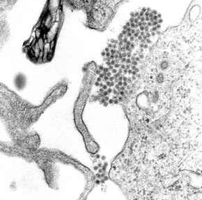
A Transmission Electron Micrograph (TM) showing dengue virus virions (the cluster of dark dots near the center).
Dengue virus is transmitted to humans through mosquitoes, and represents one of the biggest public health threats to human populations living in tropical climates (since this is where mosquitoes live). The primary symptoms of dengue include fever and severe joint pain, though more severe illness, such as dengue hemorrhagic fever or dengue shock syndrome, can occur. Over 50 million people become infected by dengue each year, with approximately 24,000 annual deaths.
Dengue is one of 50 species of viruses belonging to the Flaviviridae family of viruses. Flaviviruses are spherical in shape, and display icosahedral symmetry. Common physical features of a virion belonging to this family are a lipid bilayer envelope (complete with glycoproteins), surrounding a single-stranded RNA genome that is complexed with many copies of a small capsid protein. The outer lipid bilayer shell of a dengue virion is completely covered with exactly 180 copies (a multiple of 60!) of a glycoprotein that tightly packs in an unusual herringbone array, giving the virus a relatively smooth surface and upholding its icosahedral symmetry.
To build a paper model of a dengue virus, download the template from the Save & Share menu (or use this PDF link).
Human Immunodeficiency Virus (HIV)
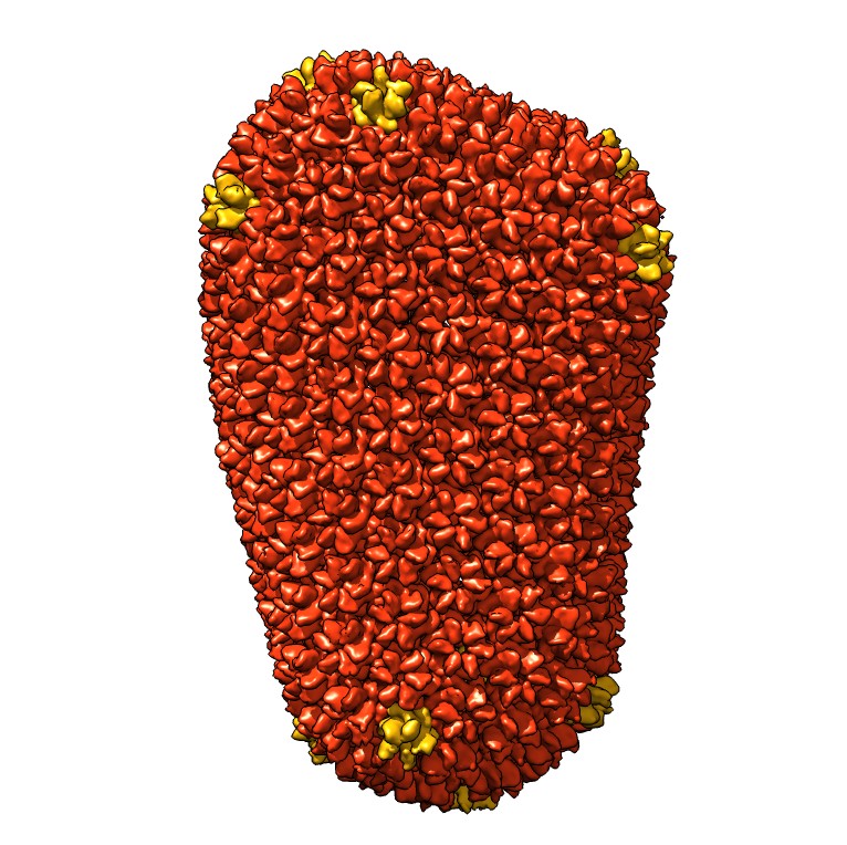
HIV Capsid Model (shared with permission from RCSB PDB).
HIV is one of the most extensively studied viruses in the world. This is attributable to its role in causing Autoimmune Deficiency Syndrome (AIDS) in humans. Over 75 million people have become infected with HIV since the beginning of the HIV epidemic in the 1980s. During this timeframe, HIV has claimed the lives of over 30 million people worldwide. As such, HIV has been at the center of social and political movements for decades.
HIV is an example of a virus that does not uphold icosahedral or helical symmetry. It has a structure characterized by a spherical lipid membrane bilayer that surrounds a capsid containing 2 copies of a single-stranded RNA genome. HIV starts off as an immature, non-infectious, spherical particle. However, as it buds off of its host cell, the HIV capsid proteins dramatically rearrange into a conical shape, forming an infectious particle, or virion. The conical HIV capsid is composed of approximately 200 copies of a single type of protein called p24. The p24 protein is quite flexible, since it is composed of two domains connected by a flexible linker. This flexibility allows the p24 protein to combine with other p24 proteins in groups of six (hexamers) or in groups of 5 (pentamers). While the HIV capsid does NOT maintain icosahedral symmetry, the fact that p24 can group into pentamers or hexamers is a prime example of quasi-equivalence, which was first proposed in the context of virus structure by Caspar and Klug in 1962 (discussed in link to symmetry thing.
To build a paper model of HIV virus, download the template from the Save & Share menu (or use this PDF link).
Zika Virus
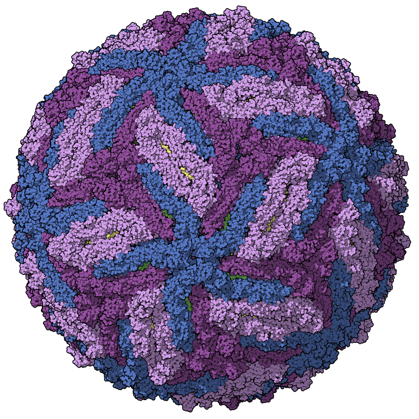
Zika Virus Capsid Model (shared with permission from RCSB PDB)
Zika virus was first discovered in 1947, when scientists isolated it from rhesus macaques living in the Zika forest of Uganda. Nevertheless, the zika virus remained in obscurity for many decades. Zika ultimately became a household name in 2015 when it made its way to the Americas through the migration of a trusty host: the mosquito. While most people who become infected with zika virus are asymptomatic, or show mild symptoms like fever or muscle pain, it does pose a significant risk for those who are pregnant. For reasons we still do not understand, zika virus can be transmitted from mother to fetus, and lead to a birth defect of the brain called microcephaly. Zika has also been associated with other pregnancy-related problems, such as miscarriage and stillbirth.
Similar to the dengue virus, zika also belongs to the Flaviviridae family of viruses. With an outward appearance that bears resemblance to a golf ball, a zika virion contains a single-stranded RNA genome encased in a capsid surrounded by a spherical lipid bilayer. Unsurprisingly, the lipid bilayer of a zika virion is packed with 180 protein units, consisting of 2 types of proteins (a membrane protein and an envelope glycoprotein). Upholding the Caspar-Klug theory, the zika virus maintains icosahedral symmetry.
To build a paper model of a zika virus, download the template from the Save & Share menu (or use this PDF link).
References
Briggs, J. A., Wilk, T., Welker, R., Kräusslich, H. G., & Fuller, S. D. (2003). Structural organization of authentic, mature HIV-1 virions and cores. The EMBO journal, 22, 1707–1715. https://doi.org/10.1093/emboj/cdg143
Centers for Disease Control and Prevention. Zika Virus Overview. https://www.cdc.gov/zika/about/overview.html
HHMI BioInteractive. Dengue virus. https://www.biointeractive.org/classroom-resources/dengue-virus
Kuhn, R. J., Zhang, W., Rossmann, M. G., Pletnev, S. V., Corver, J., Lenches, E., Jones, C. T., Mukhopadhyay, S., Chipman, P. R., Strauss, E. G., Baker, T. S., & Strauss, J. H. (2002). Structure of dengue virus: implications for flavivirus organization, maturation, and fusion. Cell, 108, 717–725. https://doi.org/10.1016/s0092-8674(02)00660-8
Ganser-Pornillos, B. K., Yeager, M., & Sundquist, W. I. (2008). The structural biology of HIV assembly. Current opinion in structural biology, 18, 203–217. https://doi.org/10.1016/j.sbi.2008.02.001
RCSB Protein Data Bank. Dengue virus. https://pdb101.rcsb.org/motm/103
RCSB Protein Data Bank. Dengue virus paper model. https://pdb101.rcsb.org/learn/paper-models/dengue-virus
RCBS Protein Data Bank. HIV capsid. https://pdb101.rcsb.org/motm/163
RCBS Protein Data Bank. HIV capsid paper model. https://pdb101.rcsb.org/learn/paper-models/hiv-capsid
RCSB Protein Data Bank. Zika virus. https://pdb101.rcsb.org/motm/197
RCSB Protein Data Bank. Zika Virus Paper Model. https://cdn.rcsb.org/pdb101/learn/resources/zika/zika-paper-model.pdf
Wen, Z., Song, H., & Ming, G. L. (2017). How does Zika virus cause microcephaly?. Genes & development, 31(9), 849–861. https://doi.org/10.1101/gad.298216.117
World Health Organization. Global Health Observatory (GHO) data. https://www.who.int/gho/hiv/en/


")


