Explore the Chemistry That Holds Us Together
Bonds are sustained chemical connections between atoms. They hold sets of atoms together in predictable proportions or even precise numbers in order to create the myriad substances with diverse physical and chemical properties we interact with each day. The strongest bonds—covalent bonds—form discrete molecules that interact with themselves and one another through weaker intermolecular forces.
These activities will explore how our familiar bio-macromolecules demonstrate the full range of bonds and intermolecular forces taught in high school chemistry and biology courses, while being engaging and relatable for students. We encourage chemistry and biology teachers talk with each other to use common vocabulary and definitions to help students apply knowledge from one class to the other, in whichever order these classes are taught.
Explore the array of resources below for various ways to apply this theme. When you like what you find, check out the full version for background information, full protocols, discussion questions, visuals, downloadables (coming soon), and more. Adapt these materials for your context, and please reach out with suggestions and ideas!
Try with your students...
Protein Denaturation and Digestion
Materials

Foods
- Unseasoned Meat Tenderizer
- Vinegar
- Milk and/or Gelatin

Lab Supplies
- Test Tubes or small beakers
- Pipettes or Measuring Spoons
- Hot plate
Procedure
Procedure For Milk Test
- Fill 6 test tubes each with 5 mL of milk.
- To each test tube containing milk, add one of the following substances:
- 1 mL water
- 1 mL vinegar
- 2 g meat tenderizer
- 1 mL meat tenderizer solution
- 1 mL boiled meat tenderizer solution
- 1 mL boiled vinegar
- Make observations immediately and over the next 30-60 minutes.
What conclusions can you draw about how acid, heat, and enzymes affect milk protein structure?
Procedure For Gelatine Test
- Dissolve gelatin in water (3 g gelatin per 100 mL water) by heating carefully on a hot plate just until dissolved. Let cool to room temperature.
- Fill 3 test tubes each with 5 mL of gelatin solution.
- To each test tube containing gelatin solution, add one of the following substances:
- 1 mL water
- 1 mL vinegar
- 2 g meat tenderizer
- Place the test tubes in an ice bath or in the refrigerator.
- Make observations immediately and over the next 30-60 minutes.
What conclusions can you draw about how acid and enzymes affect gelatin protein structure?
Single Step Lipid Extraction From Food Stuffs
Materials

Foods
Any assortment; make sure at least some contain substantial fat content. E.g.
- Ripe Avocado
- Egg Yolk
- Egg White
- Mayonnaise

Solvents / Solutions
- Chloroform*
- Methanol*
- 0.73% NaCl Solution
*Note: Chloroform and methanol should be used in a fume hood, and must be handled with the appropriate personal protective equipment, including gloves and safety goggles.

Materials for Sample Preparation
- 15mL Conical Tubes and/or Glass Tubes
- Paper Cups for manipulating food samples
Note: Lipid preparations are best done in glass since plastic materials can affect lipid extractions — glass or Teflon-coated plastics are preferred. However, this is only critical when performing analytical assays or assays that have a high sensitivity.

Equipment
- Mortar and Pestle to grind solid foods
- Spoon or Spatula
- Glass Pasteur Pipettes & Rubber Pipette Bulbs
- Rack for test tubes or similar
- Centrifuge (if you do not have a centrifuge, see note under step 7 of protocol)
- Micropipettes & Tips (if you do not have access to micropipettes, you can still perform this protocol using glass Pasteur pipettes)
Procedure
- To each tube of foodstuffs, add 1 mL of chloroform:methanol (2:1, v/v) solution.
- Secure the cap on the tube and vigorously shake the samples for 30 seconds.
- Place the tubes back into a rack, and remove the caps.
- At this point, you have created a monophasic system whereby components are dissolving in the solution.
- Add 267 μL of the 0.73% NaCl solution to each tube to make a chloroform: methanol: water mixture (2:1:0.8, v/v/v).
- Secure the caps back onto the tubes and again shake vigorously for 30 seconds.
- The addition of the aqueous salt solution transforms the mixture into a biphasic system, and helps to separate the hydrophobic lipid components from water-soluble (hydrophilic) components.
- Because chloroform is more dense than methanol and water, the chloroform-lipid phase will move toward the bottom of the tube, and on top of this will sit the less dense methanol-salt phase (thereby creating a biphasic system).
- Spin conical tubes in a benchtop centrifuge at 2,500 rpm for 2 minutes.
- Carefully remove the tubes from the centrifuge, being sure not to disturb the phase separation.
- If you do not have a benchtop centrifuge, you can allow the tubes to sit, undisturbed, for 30-60 minutes, which will allow the phases to completely separate.
- Transfer the top, methanol-water phase, to a waste container labeled for methanol disposal.
- If you have interest and ability, this phase can be used to assess non-lipid components of the food materials.
- Label a fresh glass tube with the appropriate food label.
- Using a glass pipette, transfer the bottom, chloroform-lipid phase to this freshly labeled glass tube.
- There will still be a small amount of biomass in the tube, above the chloroform-lipid phase — try to get to the chloroform-lipid phase without disturbing the biomass too much. You can just quickly “push” your glass pipette tip through the biomass to get to the bottom.
What observations can you make about the lipid samples?
- The lipid is now ready to be analyzed using TLC.
Thin Layer Chromatography (TLC) for the Separation of Lipids
Materials

Chemicals
- Lipid Extract
- Petroleum Ether*
- Diethyl Ether*
- Glacial Acetic Acid*
- Lipid Standard
- Resublimed Iodine*

Specialty Equipment
- Silica-coated TLC plates (these can be purchased from Sigma Aldrich or other similar scientific suppliers)
- Whatman Paper “wick”
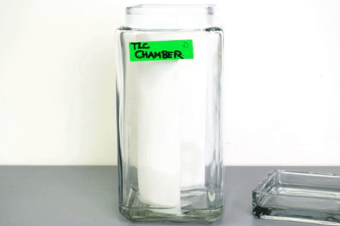
- TLC Chamber (can use ball jars)
- Pipette Tips or Glass Capillary Tubes

Lab Equipment
- Micropipette (can use glass Pasteur pipette)
- Hotplate
- Fume Hood
- Pencil
- Lab tape
Procedure
- Using 2 small pieces of tape, attach the TLC plate to the hotplate such that the bottom of the plate is touching the hotplate.
- Mark the origin (bottom) of your plate with a pencil and label each lane.
- Slowly spot the standard in the first lane in increments of 5 μL, allowing the spot to dry.
- Repeat 3 times for a total of 15 μL standard.
- Next, slowly spot the lipid extract in the designated lanes in increments of 5 μL, allowing the spots to dry.
- Repeat 5 times for a total of 25 μL sample.
- If you do not have micropipettes, you can use a glass Pasteur pipette to add your lipid extract to the plate in a dropwise fashion, being sure that you allow the “spot” to dry in between each drop
- Place the plate into the charged TLC chamber, in front of the wick.
- Secure the lid to the chamber and allow the plate to develop.
- Once the solvent front nears the top of the plate, remove the plate from the TLC chamber, quickly mark the solvent front with a pencil, and allow it to dry.
- It typically takes 30-60 minutes for the solvent front to reach near the top of the plate.
- Place the plate into the iodine chamber for visualization and secure the lid.
- Once adequately stained (about 3-5 minutes), remove and lightly outline the spots and/or take a picture.
What conclusions can you draw about the polarity of the substances?
Discuss one purpose of this technique in research or industry.


")


