Explore Microscopy Images
Take part in the microscopy image analysis process and explore more kinds of microscopes that scientists use to study various research questions. Then share your images and deeper questions in the padlet.
This work is licensed under CC BY-NC 4.0 ![]()
![]()
![]()
Preview

Analyze your own microscopy images
- Download one or more .tif files from the Save & Share Menu
- Read and/or watch the tutorial on how to use ImageJ to make a pseudocolor composite image
- Share your images on the padlet to enjoy together!

LAB Backstage Video Tour
Explore yet another type of microscopy in this LAB Backstage video tour hosted by Rockefeller University graduate student, Rochelle Shih.
Image Analysis with ImageJ
Use the image analysis software ImageJ, available in your web browser, to process some scientific images taken here at Rockefeller University.
Follow Kevin’s Tutorial
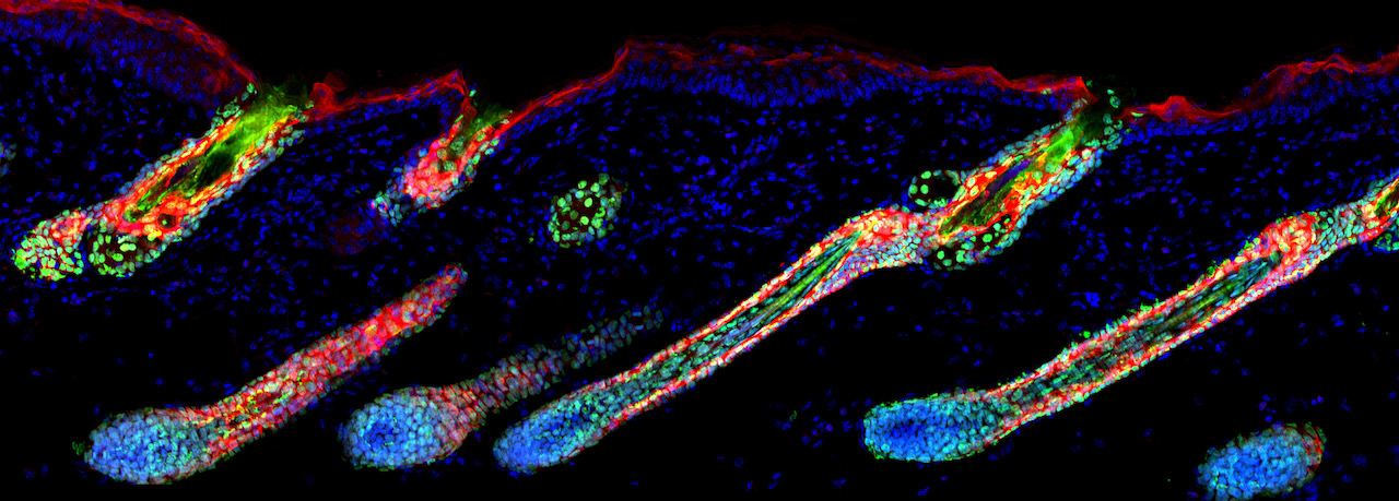
Scientist Kevin Gonzales walks us through the steps to make a pseudocolor composite image. In this case, he’s using an image he took of hair follicle cells migrating to the surface of the skin (epithelium).
Use the Save & Share Menu to download hair_follicle.tif. There you will also find a written tutorial download or follow along with the video tutorial:
Try out more images on your own
Download more images to try the same or varied techniques on different biological subjects including cells in various stages of the cell cycle and undergoing cell division. The images for this activity were collected at Rockefeller University by Megan Kelley, Lina Carlini, Donovan Phua, and Kevin Gonzales. Learn about the various images as summarized in this spreadsheet (or download it from the Save & Share Menu). Some images are available in the Save & Share Menu while others are quite large and can be downloaded from here only.
Share your beautiful images for us all to enjoy and discuss
(or share any questions if you need help troubleshooting or exploring image analysis further)
LAB Backstage Exploring Force Microscopy
Rochelle Shih is a Rockefeller University graduate student who uses a special kind of microscopy for her research. Check out her lab, and see how her microscope works in this session of LAB Backstage.


")
