Learn about microscopes
Microscopes are specialized tools that use lenses to focus energy onto objects in order to magnify them. Zoom into how microscopes work with downloadable coloring pages (including “connect the energy dots”), go on microscope tours with scientists from The Rockefeller University and BioBus, and more!
This work is licensed under CC BY-NC 4.0 ![]()
![]()
![]()
Preview

Microscope Coloring Pages
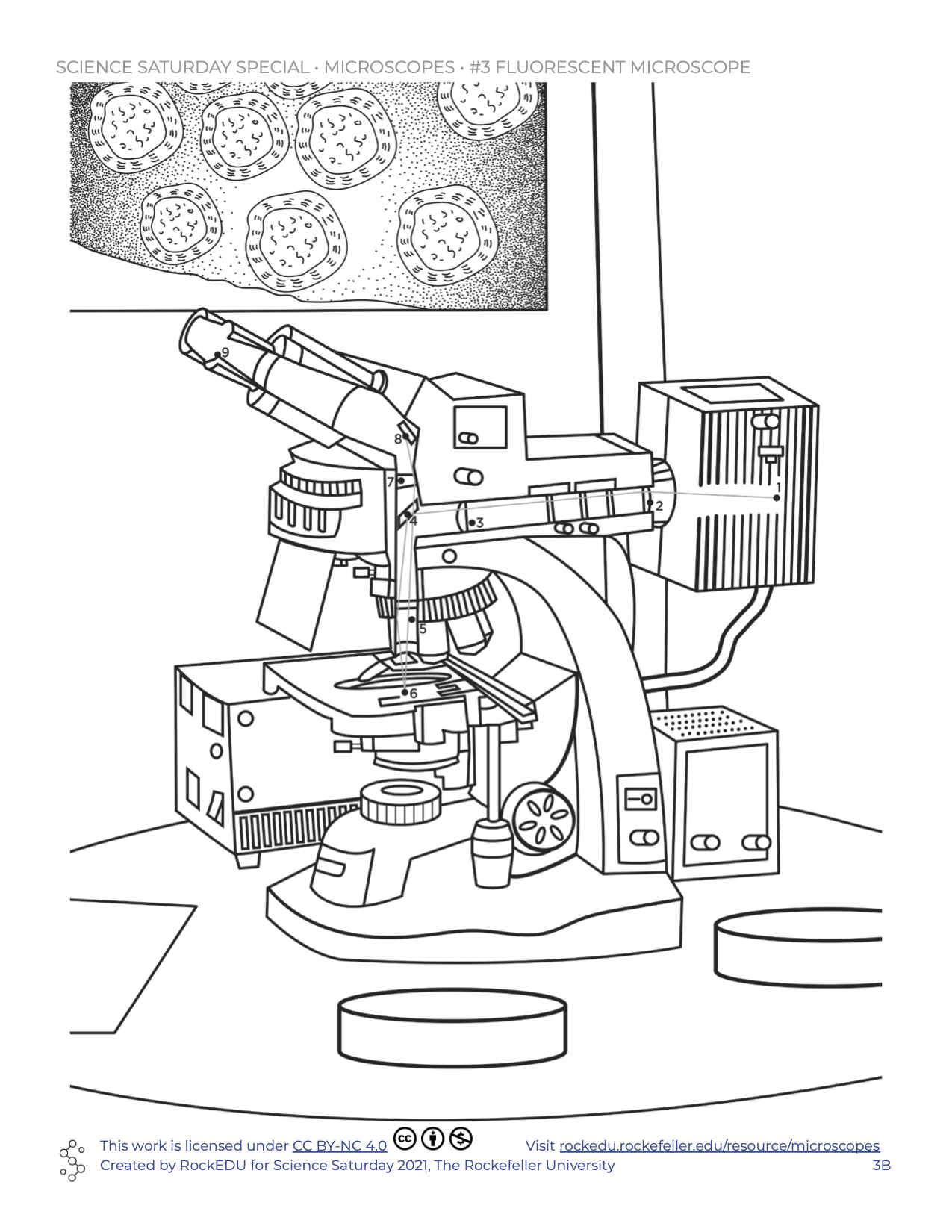
Spend some time tracing the path of illumination energy used by microscopes to magnify and focus tiny objects! These images were made by artist Jerlyn O’Donnell, aka the Design Lady.

LAB Backstage with the BioBus
RockEDU has teamed up with the amazing science education organization BioBus and 3rd graders from Massachusetts to demonstrate how a light microscope can help us see the critters in leaves that we find on the floor.

Padlet: Share Your Artwork!
Be sure to check out all the coloring pages and extra information we have in the Save & Share menu. Print out and color these pages, and share your microscopy artwork back with us!
There are many types of microscopes
There’s lots out there that are so small, they are invisible to our eyes. To help us learn about these tiny things, scientists and engineers have developed microscopes that can magnify objects up to 10,000,000 times 😲😲😲. This means that we can “see” unimaginably small things, like cells, molecules, or even atoms!
How small do you want to go?
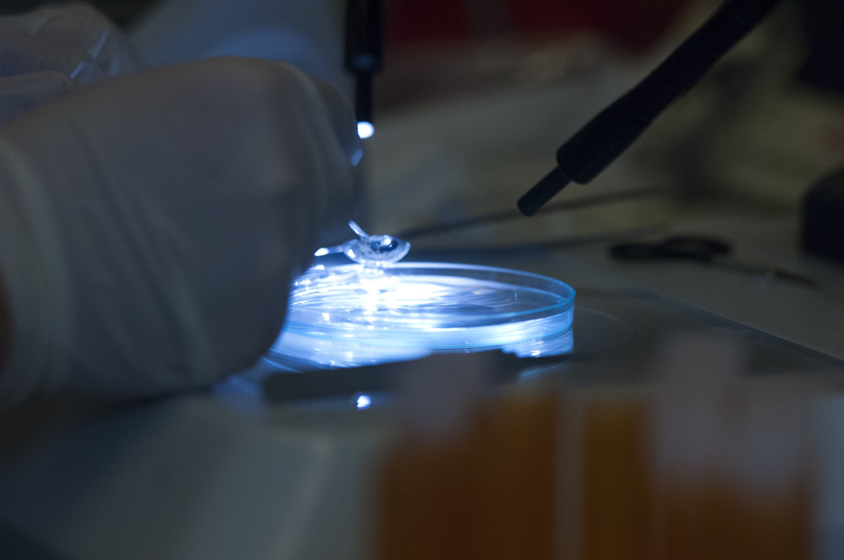
This is an essential question when determining the type of microscope that can be used. For example, have you ever seen a drop of blood and wondered “what is actually in there?” With the naked eye you may see a thick red liquid, but with different microscopes, you can see a variety of important components.
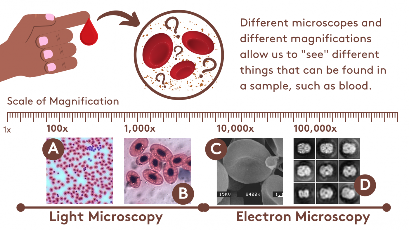
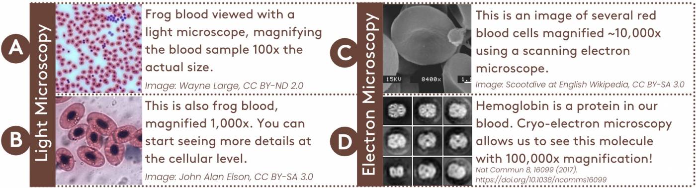 The graphic above shows examples of what we can see in blood when using two types of microscopes: light microscopes and electron microscopes. With light microscopes we can see at the level of cells and what is inside them. But with an electron microscope, the images of cells are more sharp. Additionally, an electron microscope can zoom all the way into the level of a molecule.
What is the difference between the two types of machines? To answer this question, let’s first learn about the basics of how a microscope works.
Microscope Mechanics
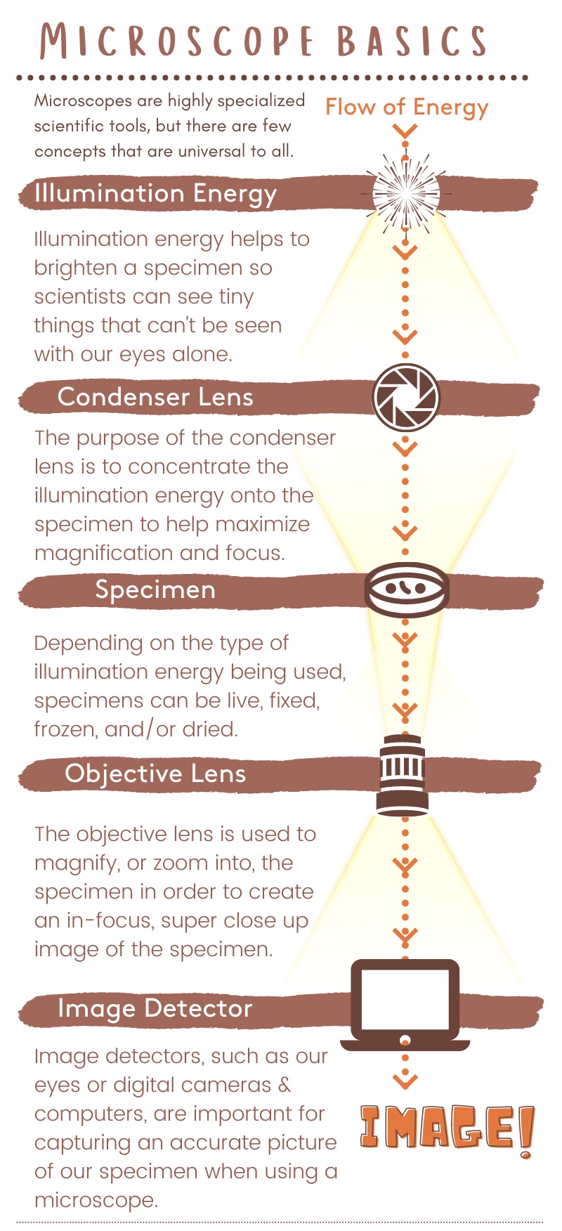
Microscopes are amazing tools with incredible and often sophisticated engineering. Below is a general schematic to demonstrate the basic principles of microscopy, or the act of using a microscope for study.
 Light microscopes have been around for several hundred years, and were first introduced to the study of biology by Dutch scientist Antonie van Leeuwenhoek in the 17th century. More recently (err, well, in 1931), scientists discovered that you can use an alternative source of illumination energy to magnify a sample: electron beams. Over the years, both the light microscope and the electron microscope have been indispensable for science.
Now, there are so many types of both light and electron microscopes. Some are easy to put together and have a small number of parts, while others require extreme environments and specialized trainings in order to build and operate them. Despite the range of complexity when it comes to the mechanics of microscopes, there are a few key parts that are common to all. This includes the concepts of illumination energy, lenses, and image detectors.
Two powerful microscopes


Both light microscopes and electron microscopes have helped scientists understand amazing things about biology, or the study of life. As you can probably guess from their category names, light microscopes work by harnessing light (spanning the electromagnetic spectrum from ultraviolet to infrared), whereas electron microscopes produce an accelerated electron beam to create illumination energy. The table above is a basic comparison of light and electron microscopes.
While the names of each type of microscope indicate the illumination energy used, the difference in materials used for the lenses might be less obvious. The purpose of a lens is to direct the illumination energy to a focal point, and in order to do this, it must be able to manipulate how the illumination energy behaves. Glass is a perfect material for manipulating light, but it is useless for manipulating electrons. Similarly, magnets are useless for manipulating light, but they work great for manipulating electrons. This is why light microscopes use glass lenses and electron microscopes use lenses made out of magnets.
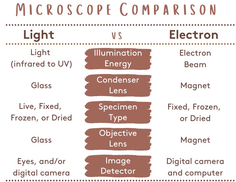
Because of the differences in illumination energy and lens type, light microscopes look completely different compared to electron microscopes. Light microscopes do not require special environments to function, and are what we typically think of when someone mentions the word “microscope.” But, because electron microscopes require super high voltages and sometimes extreme environments (such as vacuum pressure and freezing temperatures), they are generally very large.
LAB Backstage: BioBus and Leaf Litter!
Share Your Artwork With Us on Padlet
Gallery of Microscope Coloring Pages
View or download individual coloring pages here, or download the full PDF from the Save & Share menu.


")
