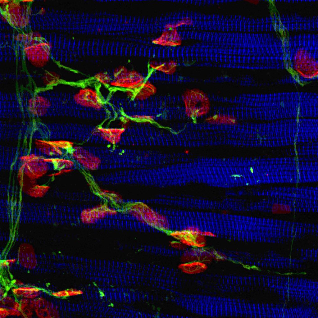
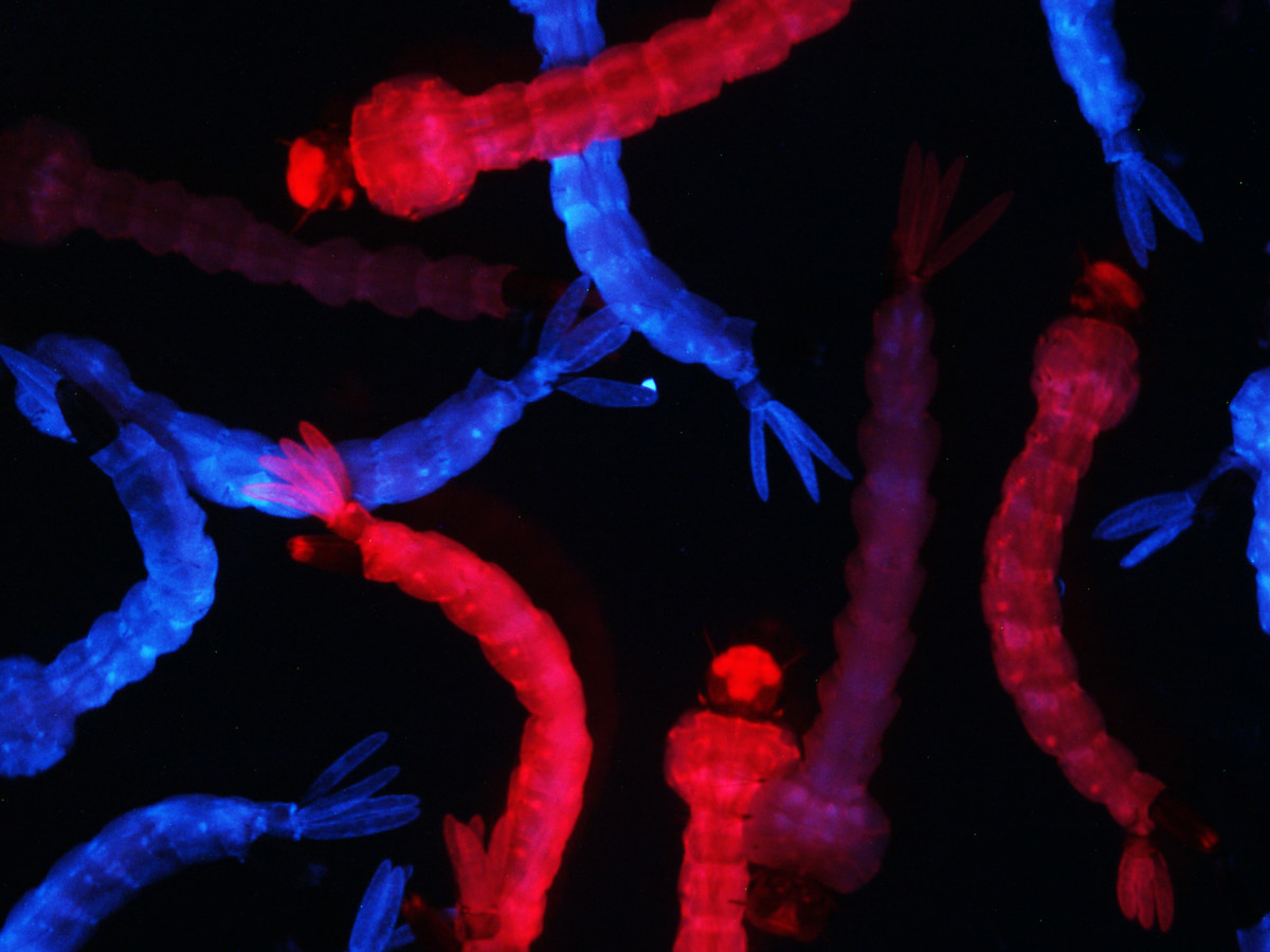
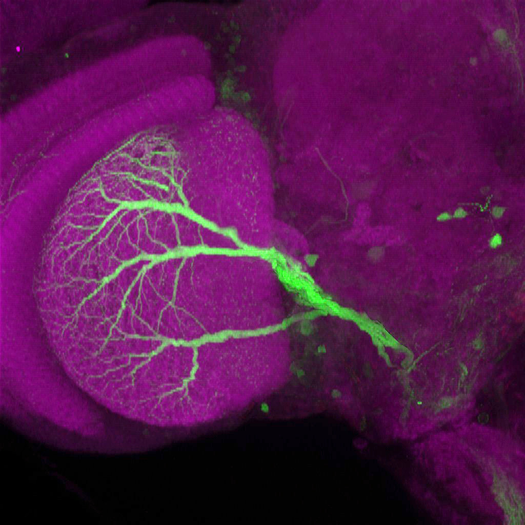
Microscopy Art Show
In this activity, kids can reassemble some beautiful microscopy images made at Rockefeller University. To do this, kids will order numbers of different kinds and with different levels of difficulty, opening up the opportunity to discuss how numbers can represent different sizes and scales for exploring our world.
Then have a scientific art show to enjoy the beautiful images!
This work is licensed under CC BY-NC 4.0 ![]()
![]()
![]()
Preview

Print & cut out the puzzle strips
Find PDFs of individual puzzles, or of the whole collection, in the Save & Share Menu.
Cut out the puzzle strips and kids can reassemble them and glue them onto the art frames to continue to enjoy their work.
Strip Puzzle Instructions
Cut out the image along all of the grey lines (making 8 strips).
Kids can order the strips based on the numbers at the bottom getting larger to reassemble the final image.
(Level up: Cut off any rows of numbers that may be easier than the kids are ready for)
Paste them into the template on the next page to make your own scientific art gallery.
Enjoy!
Images featured in these puzzles


")
